What is Nail Intramedullari and How is it Used in Surgery?
nail intramedullari is a vital technique in modern orthopedic surgery. Dr. John Smith, a renowned orthopedic surgeon, emphasizes its importance: "Nail Intramedullari revolutionizes fracture fixation." This method involves placing a rod inside the marrow cavity of a bone. It stabilizes fractures, particularly in long bones like the femur and tibia.
The simplicity of Nail Intramedullari lies in its versatility. Surgeons can use it for various injuries. Yet, it requires careful consideration. Some fractures may not heal correctly. Complications can arise, including infection or malalignment. Each case needs a unique approach, as there is no one-size-fits-all solution.
In recent years, advancements in technology have improved outcomes. However, challenges remain. Not all surgeons may be adequately trained in this technique. Ongoing education is crucial. As the field progresses, staying informed is vital for successful surgeries involving Nail Intramedullari.
Definition of Nail Intramedullari: Key Concepts and Terminology
Nail intramedullari refers to a specific surgical technique. It involves placing a metal rod inside the medullary cavity of a long bone. This method is commonly used to stabilize fractures, especially in long bones like the femur or tibia. Surgeons select this approach to facilitate healing and maintain the bone's alignment.
Key concepts in nail intramedullari include stability and support. The intramedullary nail is designed to distribute load evenly across the bone. This can enhance the healing process. The procedure usually requires careful planning and imaging. Surgeons must assess the fracture type and bone quality. Challenges can arise, such as poor bone density or alignment issues.
Terminology is important in understanding this technique. Terms like reaming or locking screws are often used. Reaming prepares the bone for the nail. Locking screws secure the nail in place. This terminology might seem complicated, but each term has a specific role. As with any surgical procedure, complications can occur. Understanding these terms helps clarify the process and improve communication between medical professionals and patients.
Historical Development of Intramedullary Nails in Surgical Practice
The history of intramedullary nails is fascinating. Developed as a solution for fractures, they revolutionized orthopedic surgery. The concept dates back to the early 20th century. Surgeons began exploring internal fixation methods. Early attempts faced numerous challenges. The materials used were often inadequate.
In the 1940s, significant advancements took place. Surgeons began to use metal rods for stabilization. These rods were inserted into the medullary cavity of the bone. This technique reduced the need for external casts. As time progressed, surgical methods evolved.
The design of intramedullary nails improved, leading to better patient outcomes.
Tips: When considering surgery, consult experienced surgeons. They can provide insights based on your unique situation. Be open to discussing potential risks. Patient education is crucial. Understand what the procedure involves and what to expect during recovery. Always ask questions about the process.
Indications for Using Nail Intramedullari in Orthopedic Surgery
Nail intramedullari, commonly known as intramedullary nailing, is a surgical technique mainly used for fractures in long bones. This method has become a preferred choice in orthopedic surgery due to its minimally invasive nature. Surgeons implant a metal rod into the medullary canal of the fractured bone, facilitating stability during the healing process.
Indications for using nail intramedullari often include diaphyseal fractures, which are mid-shaft breaks in long bones like the femur or tibia. These fractures typically result from high-energy trauma, such as accidents. In some cases, it is applicable for complex fractures with multiple fragments. The procedure can reduce recovery time significantly. Yet, complications may arise, such as infection or improper alignment.
Despite the advantages, there are reflective points to consider. Not all fractures are suitable for this method. Surgeons must evaluate each case individually. Some patients may have pre-existing conditions that complicate the surgery. Additionally, the type of nail selected and the technique's execution play critical roles in patient outcomes. Overall, while intramedullary nailing offers promising results, a careful assessment of its suitability is essential.
Nail Intramedullari Usage in Orthopedic Surgery
Surgical Techniques Involved in Intramedullary Nail Placement
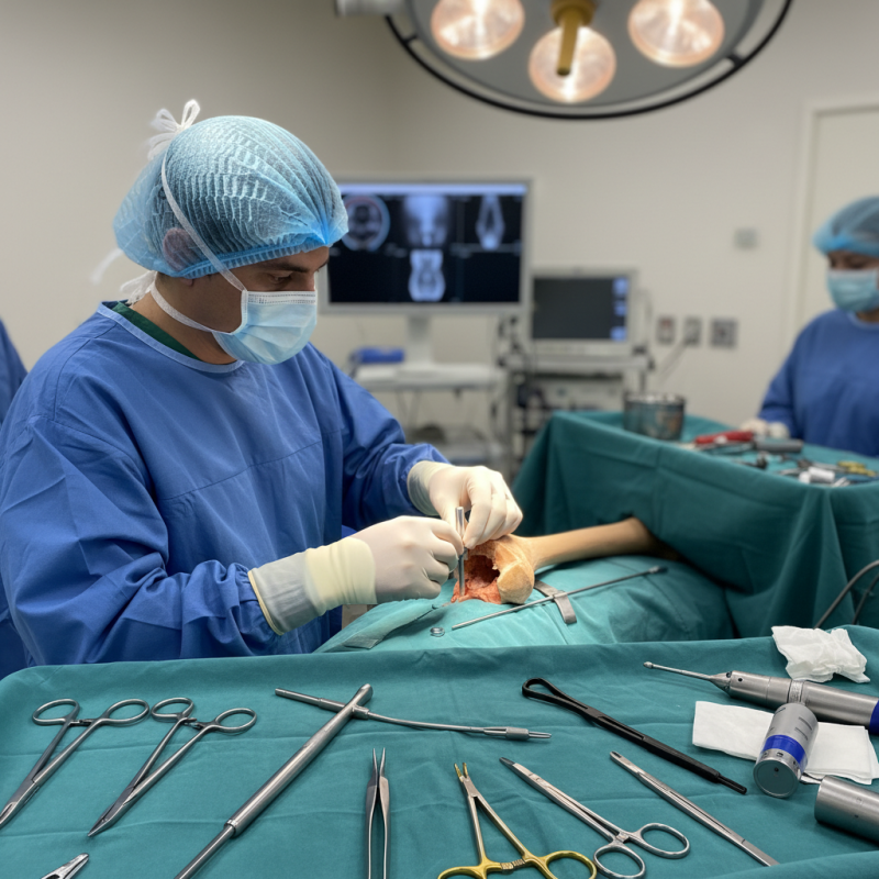
Intramedullary nailing is a common surgical technique used to treat fractures, especially in long bones. The process usually involves inserting a metal rod into the medullary canal of the bone. This technique can stabilize the fracture and promote healing. However, the placement of intramedullary nails is not without challenges.
Surgeons must consider various factors before proceeding. Proper alignment of the nail is crucial to ensure the best outcomes. The type of fracture, patient anatomy, and specific injury are all essential considerations. Some surgeons may struggle with achieving optimal placement. Misalignment can lead to complications such as improper healing or further injury.
There are also techniques that improve precision. Fluoroscopy is often used to guide nail placement accurately. This imaging technique allows real-time visualization of the bone during the procedure. Additionally, experienced surgeons often rely on their tactile feedback when inserting the nail. However, even skilled surgeons can face difficulties. Adjustments may be necessary mid-procedure, which can prolong surgery and impact patient recovery. This highlights the importance of continuous learning in surgical techniques.
What is Nail Intramedullari and How is it Used in Surgery? - Surgical Techniques Involved in Intramedullary Nail Placement
| Aspect | Details |
| Definition | Intramedullary nail is a surgical device used to stabilize fractures of long bones by being placed within the medullary cavity. |
| Indications | Used primarily for diaphyseal fractures of the femur, tibia, and humerus. |
| Advantages | Allows for early mobilization, minimal soft tissue dissection, and preserves blood supply to the fracture site. |
| Surgical Technique | Includes reaming of the medullary canal, inserting the nail, and securing it with locking screws. |
| Complications | Potential complications include infection, malunion or nonunion of the fracture, and damage to surrounding structures. |
| Post-operative Care | Includes pain management, physical therapy, and follow-up evaluations for healing. |
Postoperative Care and Rehabilitation Following Intramedullary Nail Surgery
Postoperative care after intramedullary nail surgery is crucial for optimal recovery. Complications can arise if proper care is neglected. Studies suggest that about 10% of patients may experience postoperative issues. These can include infection and delayed healing. Pain management is a priority during this period. Patients should follow their surgeon's instructions for medication closely.
Rehabilitation often starts within days after surgery. Physical therapy plays an essential role in restoring movement and strength. A recent report noted that timely rehabilitation can improve outcomes by 30%. Exercises may focus on range of motion and gradually increasing weight-bearing activities. However, individual recovery varies, and some may struggle with progress. It’s important to maintain an open dialogue with healthcare providers regarding any concerns.
Tips: Maintain a daily log of your pain levels and mobility. This can help you track improvements and communicate effectively with your physical therapist. Don't rush the recovery process. Understand that each person heals at their own pace. Engage in light activities as tolerated, but listen to your body. This could prevent setbacks.